
Articulators
Three-dimensional movement for functional prosthetics
Design

0/0
Design
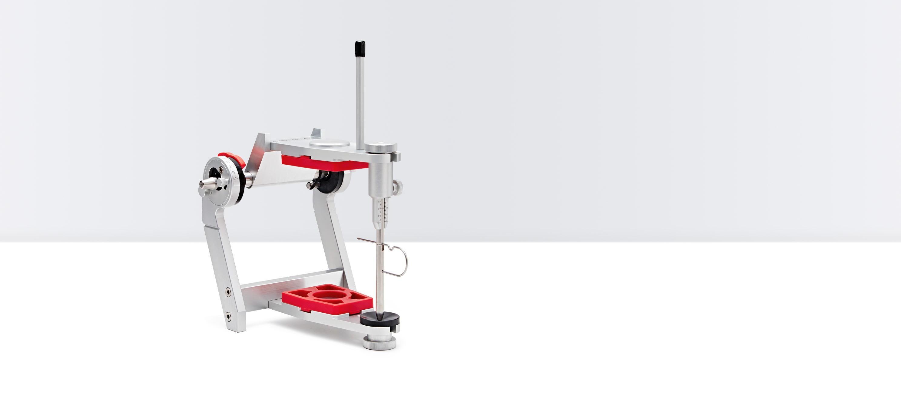
Handy, light-weight and robust aluminum construction. This design facilitates simple, physiological operation procedures. Heavily stressed elements such as the joint and vertical pin are made of stainless steel. The incisal plate is made of durable POM acrylic.
1 Incisal pointer CA 3.0
2 Retention discs
3 Plastic mounting plates
4 Incisal guide standard CA 3.0
5 Vertical guide rod CA 3.0
6 Vertical guide rod holder CA 3.0
7 Supporting rod for opening stop
8 O-ring seals
9 Knurled thumb screw A
10 Knurled thumb screw B
Technical data

0/0
Technical data
- Height: 150 mm
- Width: 145 mm
- Weight: 690 g
- Inner construction height: 100 mm (with Split-Cast)
- Depth: 160 mm
- Bonwill triangle: 110 mm
- Balkwill angle: 25°
- Retrusion track: 1.5 mm
- Immediate side-shift stop: 0 – 2.5 mm fix
- Angle of jaw track adjustable between 0° – 60°
- Incisal plate: 15°
- Material: anodized aluminum
Synchronization

0/0
Synchronization
Together with the Plate Set for Splitex® and the Calibration Key compatible with Splitex®", the CA 3.0 LARGE can be adjusted to the overall height of 126 mm and synchronized with the Artex® articulators of the Carbon series from Amann Girrbach.
«Splitex® and Artex® are registered trademarks of Amann Girrbach GmbH, 75177 Pforzheim, DE»
Technical data

0/0
Technical data
- Height: 160 mm
- Width: 145 mm
- Weight: 735 g
- Inner construction height: 126 mm (with Split-Cast)
- Depth: 160 mm
- Bonwill triangle: 110 mm
- Balkwill angle: 25°
- Retrusion track: 1.5 mm
- Immediate side-shift stop: 0 – 2.5 mm fix
- Angle of jaw track adjustable between 0° – 60°
- Incisal plate: 15°
- Material: anodized aluminum
Plate set for use with Splitex®

0/0
Plate set for use with Splitex®
- Can be integrated with CAD/CAM scanner
- Adjustment can be made with the Candulor centering key; but also with an existing Splitex®-based centering key
Compatible centering key for Splitex®

0/0
Compatible centering key for Splitex®
- Split centering key
- For the synchronization of several articulators with each other
An overview

0/0
An overview
The temporomandibular joint is both a rotating and sliding joint with two types of movement: the rotation or hinged axis as well as the translational or sliding movement. The most common form of movement is a combination of both types of movement. A rotation with simultaneous translation – a rotary sliding. Interaction between the two temporomandibular joints allows rotational movement along axes other than the hinged axis. Three axes of movement can be determined.
- A horizontal-transversal joint axis as simple hinged axis (green).
- A vertical joint axis for the respective condyle on the side of lateral movement (blue). The condyle on the opposite side performs a downward and sliding movement forwards.
- A horizontal-sagittal joint axis (black) for the respective condyle on the side of lateral movement, with simultaneous lateral movement of the joint head of the opposite side downwards and forwards.
Simulating natural movement patterns

0/0
Simulating natural movement patterns
The articulator series excels through its simple handling and precision. It was designed along the camper plane. It runs parallel to the occlusion plane, thus giving unambiguous and practical reference for daily work.
The articulator joint is analog to the natural temporomandibular joint

0/0
The articulator joint is analog to the natural temporomandibular joint
The shape of the natural joint head is copied and simulated by the double cone in the Candulor articulators. This avoids unphysiological, straight movement patterns in lateral and transverse motion sequences.
Simulating natural movement

0/0
Simulating natural movement
The articulator series is equipped with two guide edges which guide the double cone.
A vertical-sagittal one on the retrusion bar (red) and a horizontal-sagittal one on the joint disk (green).
The vertical-sagittal edge on the retrusion bar touches the inner cone (red) whereas the horizontal-sagittal edge guides along the apex of the double cone (green).
As a result, with lateral movements, the double cone is not only guided into a physiological, three-dimensional movement via the horizontal guide edge of the joint disk (green), but also via the vertical guide edge on the retrusion bar (red).
The Bennett movement

0/0
The Bennett movement
The condyle moving on the non-working side is the mediotrusion condyle: during a lateral movement (mediotrusion) the moving condyle moves forward in downward direction.
The condyle resting on the working side is the laterotrusion condyle: this side is used for chewing and the teeth are in occlusion. This condyle can rotate around the vertical axis and also move laterally.
Both articulators allow virtually identical natural 3D simulation of the lateral and Bennett movement. Functional hyper-contacts are avoided.
Sandra Räuber
CANDULOR AG
Simply send us your contact data – we will contact you promptly.


